The Quest
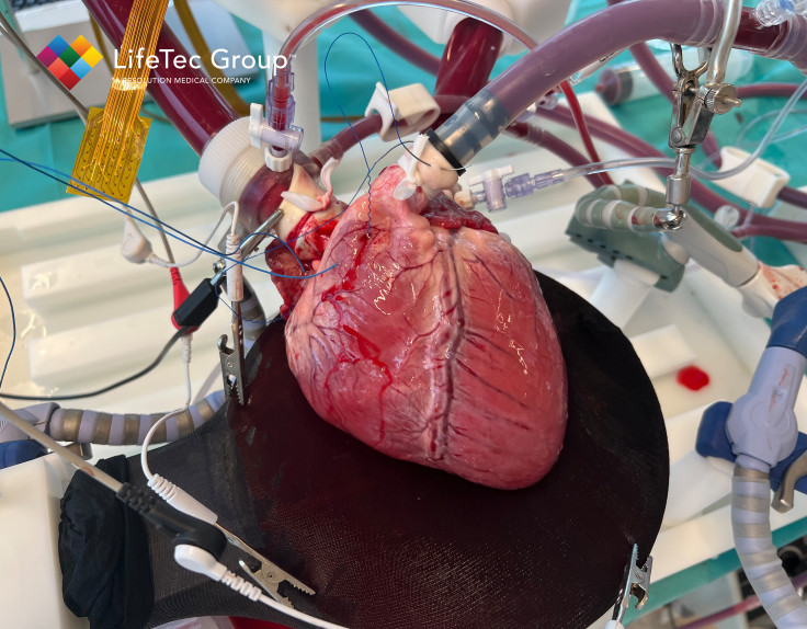
LifeTec Group’s PhysioHeart is an ex vivo beating heart platform based on abattoir tissues, a cardiac simulator that has been used for many years in the preclinical research domain. The PhysioHeart can provide useful insights because it is living and contracting, under physiological pressures and flows. So far, the PhysioHeart has mostly been used for structural interventions that were focussing on hemodynamic responses. Besides structural interventions, the platform has proven itself for imaging studies as well for most of the common clinical modalities you can think of: Ultrasound, CT, MRI, Fluoroscopy and intracardiac videoscopy. Despite these many years of research, there is another area where a lot can be learned still: the electrophysiological behaviour of the heart, especially in the atria.
The Project
Within SIGNET, (Sensing and Image-Guided Neurological therapies, cardiac Electrophysiology and Tumour treatments), the overall objective is to develop efficient MR based image-guided treatment workflows to replace currently complex procedures in cardiology, oncology, and neurology. The goal is to create single-episode, personalized, dose-adaptive, high-precision Magnetic Resonance guided treatments and interventions. These developments are aimed to improve patient comfort, safety and treatment outcome but also be more economically viable and be less dependent on staff availability.
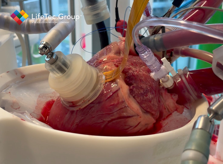
LifeTec Group is involved in the cardiac use case: treatment of cardiac arrythmia by ablation. Cardiac arrythmia occurs when the natural electrical conductance pathway of the heart is disturbed. This results in unsynchronized contractions of the heart that lead to an inefficient heart function. The disturbance of the conductance pathway often originates at the ostia of the pulmonary veins. During ablation, tiny scars are created to block these abnormal electrical signals by advancing a catheter in the groin, up to right atrium, cross the atrial septum and then reach the target area. However, before a patient can finally receive this treatment, several hospital visits are required, multi-modality imaging and planning are required and the procedure itself can be challenging.
In SIGNET we aim to develop MR guided Electrophysiology ablation (MRgEP). During ablation procedures swelling occurs which also blocks electrical signals such that the procedure looks effective. When the swelling resolves after a couple of days, the arrythmia’s return. One main advantage of using MR as a guidance technique is the ability to distinguish between scarred tissue and swollen tissue. And what better object could be used to test such technique then LifeTec Group’s PhysioHeart?
Cardiac Ablation
LifeTec Group™ is mainly involved in the cardiac use case: treatment of cardiac arrythmia by ablation. Cardiac arrythmia occurs when the natural electrical conductance pathway of the heart is disturbed. This results in unsynchronized contractions of the heart that lead to an inefficient heart function. The disturbance of the conductance pathway often originates at the ostia of the pulmonary veins. During ablation, tiny scars are created to block these abnormal electrical signals by advancing a catheter in the groin, up to right atrium, cross the atrial septum and then reach the target area. However, before a patient can finally receive this treatment, several hospital visits are required, multi-modality imaging and planning are required and the procedure itself can be challenging.
In SIGNET we aim to develop MR guided Electrophysiology ablation (MRgEP). During ablation procedures, besides the desired scar creation also swelling of the treated tissue occurs and this also blocks electrical signals. In some cases, the ablation procedure initially seems to have been effective, but when the tissue swelling resolves after a couple of days, the arrythmia’s return. One main advantage of using MR as a guidance technique is the ability to distinguish between scarred tissue and swollen tissue. And what better object could be used to test such a technique than LifeTec Group’s PhysioHeart?
What will we do?
That is right, in the 3 years of this project, we will transform the PhysioHeart to an MR-compatible testing phantom to evaluate ablation therapy effectiveness. However, the electrophysiological behavior of the atria in the Physioheart platform has not been researched before. This will be the start of the project: learn about the electrophysiology of the PhysioHeart platform and try to perform mapping measurements on it. Once that is achieved in our lab in Eindhoven and the healthy/reference status of the platform is known, we will transform this into an MR-compatible version and bring it to the MRI where real ablation studies will be performed.
Results so far
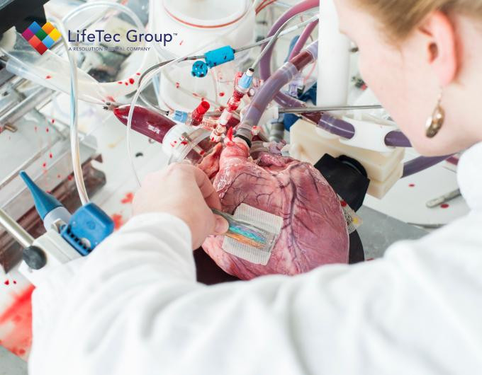
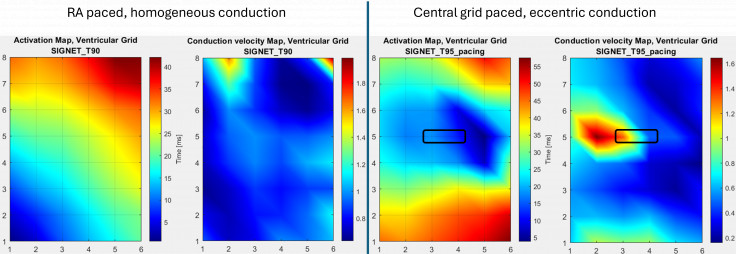
Since the start of the SIGNET project, we have been creating our own electrode grids that can be placed on the epicardium to measure electrograms. After multiple revisions, we are now working with a 6x8 grid that can be used to visualize activation patterns, like what is used in clinic. The image below shows a typical activation map. If for example arrythmias occur, such maps can visualize how and where these abnormalities originate.
To improve even further on this way of visualization, we started creating voltage maps as well. The same electrode grid is being used to generate the data, but instead of plotting just the activation timepoints in one graph, we plot the voltage amplitude of each signal over time, which gives an even better understanding of the wave propagation.
For the next step in the development of this technology, we would like to get a sense of how well we are able to detect abnormal conduction patterns such as re-entries. To mimic such a ventricular re-entry arrhythmia we conducted an experiment on our PhysioHeart platform. A porcine heart was harvested, revived and tuned to physiologic haemodynamics. Initially pacing was applied in the right atrium (RA), targeting the SA-node. Activation and conduction velocity maps were acquired using our 6x8 electrode grid on the left ventricle parallel to the LAD, showing a homogeneous pattern (see images on the left). Next, we implemented a switch connecting two central electrodes ([5,3] + [5,4]) to our pacing device instead of the RA pacing lead. At this time the heart was being paced centrally in the electrode grid (indicated by the black rectangle in images on the right) on the same left ventricle position parallel to the LAD. We acquired the electrical signals again using the grid showing an eccentric propagation pattern in line with our expectations! This proves that we are able to detect local stimuli and altered propagation patterns which can be of great value for any device that affects the electrophysiological behaviour of the heart, e.g. ablation and pacing studies!
What's in it for you?
If you are working in MR-guided interventional therapies or in cardiac electrophysiology therapies, we will develop great opportunities for you to investigate your new treatment concepts. The compatibility of the PhysioHeart with MR imaging will be greatly improved, which will become accessible to your project as a research platform. Moreover, a lot of knowledge on the electrophysiological behaviour of our ex-vivo model and interventions to influence this behaviour will be generated and this will open new opportunities for you to work with us. Are you looking for a cost-effective, cruelty-free, time-efficient, and controlled way to evaluate the performance of your ablation or pacing device?