The Study
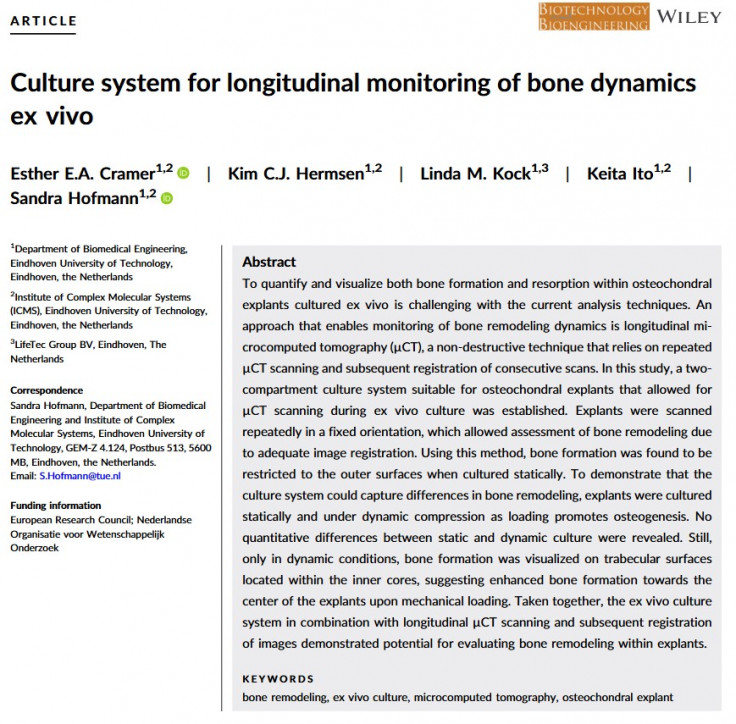
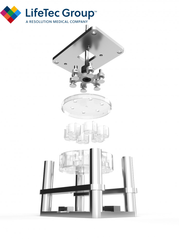
Gaining insight into bone remodeling processes could enable osteochondral cultures to be utilized as model to study novel therapies that affect formation and/or resorption processes and diseases affecting both tissues. Therefore, this study aims to establish a two‐compartment culture system for osteochondral explants that is compatible with µCT scanning thereby enabling longitudinal monitoring of bone formation and resorption during culture. Without the need to take samples out of the culture system before each scan, it is expected that ensuring a similar position during each scan will reduce the risk of errors and misalignment during registration, thereby enabling registration of the complete volume of the explant. To demonstrate that the culture system could capture differences in bone turnover, in the second part of this study we aimed to implement compressive mechanical loading to enhance bone formation ex vivo. To ensure imposed compressive forces are received by the bone tissue, bone explants without a cartilage layer were utilized, because cartilage will alter loading patterns experienced by the bone. This makes culture in a two‐compartment system unnecessary. Hence, our culture system was designed to be adjustable for scanning of bone explants also in one compartment and thereby allows for monitoring of bone dynamics in both osteochondral and bone explants.

Results and conclusion
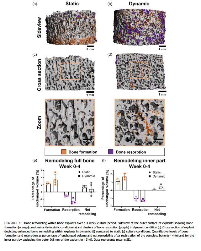
Interestingly, both statically and dynamically cultured bone explants showed significantly higher levels of cell death during the first 4 days of culture, which stabilized towards low levels from Day 7 onwards. Moreover, after registration of week 4 µCT images onto baseline images, it was observed that dynamically cultured samples showed increased amounts of formation in central areas of the bone explants.
In conclusion, this culture system, when osteoclast activity could be implemented, has the potential to be a platform for standardized ex vivo evaluation of novel bone‐related therapeutics, including materials for bone defects as well as bioactive agents for bone diseases.

What's in it for you
The research studies presented in this paper is a typical example of collaborations in which LifeTec Group loves to engage. This study nicely showcases one of the features of the osteochondral platform, the dynamic loading part. The platform is the translation of recognized fundamental science into applied science: it provides you exactly that lifelike ex-vivo setting for testing your product, at the same time reducing the amount of animal testing needed. If you are working on medical devices, tissue regeneration or pharmacological therapies, a collaboration project with our team allows you to fully focus on the device or therapy development and LifeTec Group will act as an extension to your team to focus on the environment that allows us to demonstrate together if the device or therapy is promising. And together we can speed up the process: by applying realistic tissue environments, we can learn a lot in relatively short time before needing to proceed to the necessary but ethically burdened animal tests that will deliver the "Go" for human trials.
If you're interested to learn how LifeTec Group can support your project, then feel free to review our platforms and services, example cases and contributions to scientific publications that you will find on our website, and please reach out to our team for more information and backgrounds.

Interested in more about what we do at LifeTec Group? Contact us!
Call at +31 40 2989393 Or e-mail us
