Study / Scientific Publication
Keywords: Full-field measurements, Three-dimensional digital image correlation (3D-DIC), Epicardial strain, In vitro porcine heart, Repeatability, High-resolution | Cardiac Biosimulator Platform
Cardiac strain imaging
The quantitative assessment of cardiac strain is increasingly performed to provide valuable insights on heart function.
Currently, the most frequently used technique in the clinic is ultrasound-based speckle tracking echocardiography (STE).
However, verification and validation of this modality are still under investigation and further reference measurements are required to support this activity

Study
The aim of this study was to enable these reference measurements using a dynamic beating heart simulator to ensure reproducible, controlled, and realistic haemodynamic conditions and to validate the reliability of optical- based three-dimensional digital image correlation (3D-DIC) for a dynamic full-field analysis of epicardial strain.

Abstract
Specifically, performance assessment of 3D-DIC was carried out by evaluating the accuracy and repeatability of the strain measurements across multiple cardiac cycles in a single heart and between five hearts.
Moreover, the ability of this optical method to differentiate strain variations when different haemodynamic conditions were imposed in the same heart was examined.

Strain measurements were successfully accomplished in a region of the lateral left ventricle surface. Results were highly repeatable over heartbeats and across hearts (intraclass correlation coefficient = 0.99), whilst strain magnitude was significantly different between hearts, due to change in anatomy and wall thickness. Within an individual heart, strain variations between different haemodynamic scenarios were greater than the estimated error of the measurement technique.
This study demonstrated the feasibility of applying 3D-DIC in a dynamic passive heart simulator. Most importantly, non-contact measurements were obtained at a high spatial resolution (~ 1.5 mm) allowing resolution of local variation of strain on the epicardial surface during ventricular filling. The experimental framework developed in this paper provides detailed measurement of cardiac strains under controlled conditions, as a reference for validation of clinical cardiac strain imaging modalities.
Results:
- The 3D-DIC analysis was successfully accomplished in all the hearts over a region of the left ventricle surface, however, the reconstructed ROIs were slightly different between hearts because of changes in the anatomical structures and positioning within the cameras FOV.
- The strain errors obtained between the five successive reference states of the heart (unloaded configuration) were less than 0.2% in all cases. The median value and range of the average and standard de- viation of the surface strain values in the five hearts are reported in Table 2.
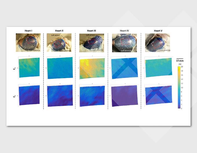
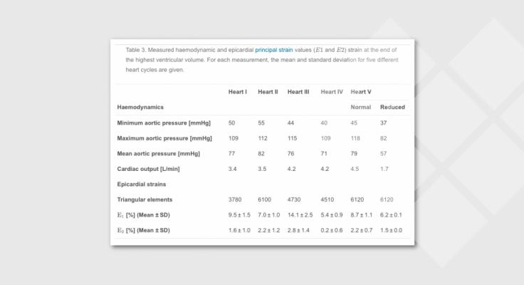
- The repeatability assessment reported here was performed under the measured haemodynamic parameters reported in Table 3. These were selected for each heart to be within a physiological range. 3D-DIC repeatability assessment was described both quantitatively through the ICC and qualitatively by plotting the average of principal strain surface values over the cardiac cycles, as shown in Fig. 4. The ICC showed an excellent repeatability of 3D-DIC in assessing strains with values of 0.99 for a CI of 95% in both E1 and E2.
- As shown in Fig. 4, the frequency of the temporal strain variation in each heart was very similar to the heart rate applied to the pump (70 bpm). Starting from near zero, average strains increased in magnitude as the ventricle expanded and returned to zero at end of the cardiac cycle. The number of mesh triangular elements used in the strain volume and is shown in Fig. 5, along with the minimum principal strain directions, orthogonal to the maximum principal strain directions. The behaviour of the strain distribution throughout the cardiac cycles can be observed in the videos in the Supplementary Materials.
Supplementary material related to this article can be found online at doi:10.1016/j.jmbbm.2018.11.025. - To demonstrate whether 3D-DIC was able to differentiate haemo- dynamic changes such as a hypotensive state of the heart (Leopaldi et al., 2015), in one heart (Heart V) the mean aortic pressure was ad- justed to 57 mmHg. The strain distribution in the same heart under these different haemodynamic conditions are illustrated in Fig. 6, showing variation in peak strain between the two states of around 2.5%.
-
You can download this paper below, or visit the ScienceDirect webportal [link]
Fig. 1
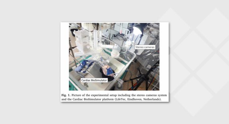
Fig. 2
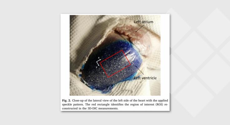
Fig. 3
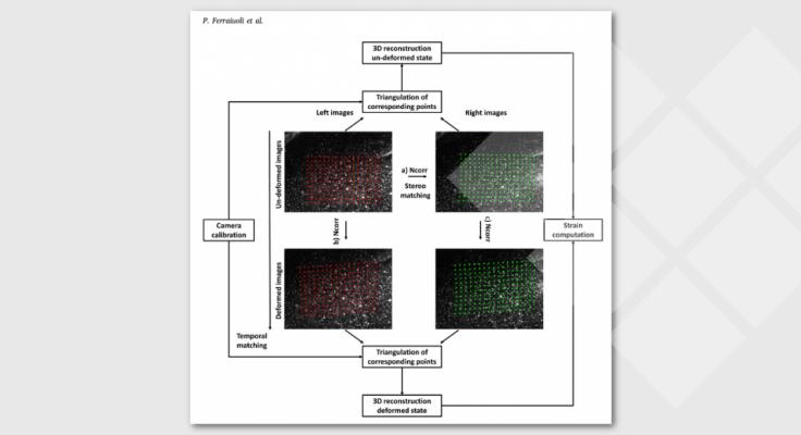
Fig. 4
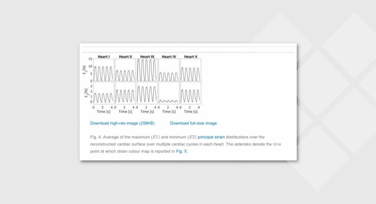
Fig. 5
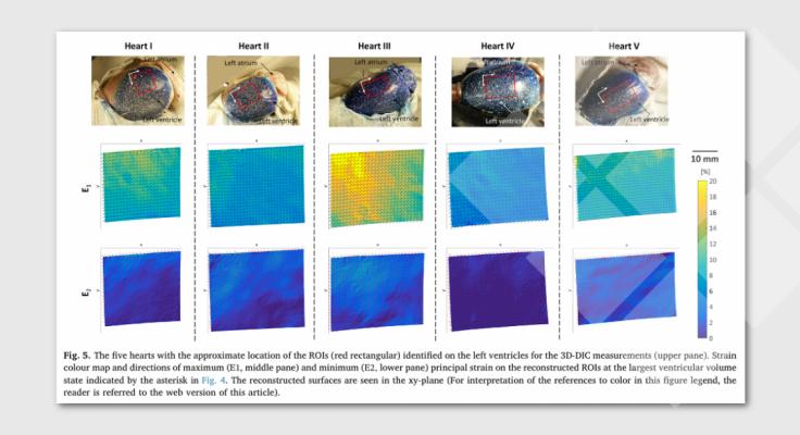
Fig. 6

Table 1
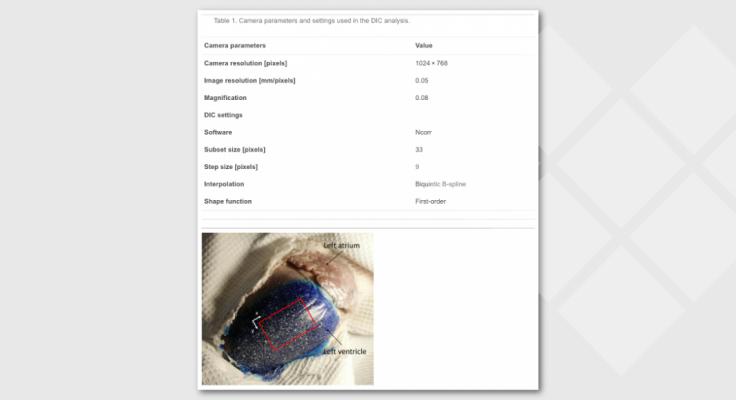
Table 2

Table 3
Authors:
Paolo Ferraiuoli, Benjamin Kappler*, Sjoerd van Tuijl*, Marco Stijnen*, Bas A.J.M. de Mol*, John W. Fenner, Andrew J. Narracott
- Mathematical Modelling in Medicine Group, Department of Infection, Immunity and Cardiovascular Disease, University of Sheffield, Sheffield, UK
- Insigneo Institute for in Silico Medicine, University of Sheffield, Sheffield, UK
- (*) LifeTec Group, Eindhoven, Netherlands
- Department of Cardiothoracic Surgery, Academic Medical Center, Amsterdam, Netherlands
This work was funded by the European Commission, Belgium through the H2020 Marie Skłodowska-Curie European VPH-CaSE Training Network (www.vph-case.eu). GA No. 642612.
- COLOFON
Acknowledgements

Interested in more about what we do at LifeTec Group? Contact us!
Call at +31 40 2989393 Or e-mail us
